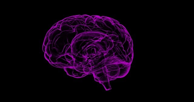
Advanced medical imaging techniques like MRI and CT have significantly enhanced the assessment and diagnosis of spinal cord injuries, providing detailed insights into the nervous system through non-invasive methods. Techniques such as DTI allow healthcare professionals to accurately assess axonal damage and predict outcomes, revolutionizing treatment planning for nervous system injuries. These technologies are pivotal for prompt intervention and improved patient care in medical imaging for the nervous system.
Advanced imaging techniques are transforming the way we detect and diagnose spinal cord injuries, revolutionizing care in the field of neural trauma. This article delves into the cutting-edge tools that enable precise assessment of the spine and nervous system. We explore medical imaging’s crucial role in identifying damage to the delicate structures within the spinal cord, with a focus on non-invasive scanning methods. By harnessing these modern techniques, healthcare professionals enhance diagnosis, offering improved outcomes for patients suffering from these potentially debilitating injuries.
Advanced Imaging Techniques for Spinal Cord Assessment
Advanced imaging techniques have revolutionized the assessment of spinal cord injuries, offering unprecedented insights into this critical region of the nervous system. Techniques such as magnetic resonance imaging (MRI) and computed tomography (CT) scans provide detailed cross-sectional images, enabling healthcare professionals to detect and diagnose spinal cord damage with greater accuracy.
MRI, in particular, excels in visualizing soft tissues, making it invaluable for identifying subtle changes in the spinal cord’s structure and function. Diffusion tensor imaging (DTI), a specialized MRI technique, tracks the movement of water molecules within neural fibers, providing information on white matter integrity. This capability is crucial for assessing the extent of axonal damage and predicting functional outcomes in spinal cord injuries.
Role of Medical Imaging in Nervous System Trauma Detection
Medical imaging plays a pivotal role in detecting and diagnosing spinal cord injuries, offering crucial insights into the extent of nervous system trauma. These advanced techniques allow healthcare professionals to visualize internal structures with remarkable detail, enabling them to identify even subtle damage that might be missed through traditional examination methods. By employing technologies such as magnetic resonance imaging (MRI) and computed tomography (CT), doctors can non-invasively assess the spine, brain, and surrounding tissues, providing critical information for accurate diagnosis and treatment planning.
In cases of suspected spinal cord injury, medical imaging provides a comprehensive view, helping to detect hematomas, fractures, disc herniations, or other structural abnormalities. MRI, with its ability to generate detailed cross-sectional images, is particularly valuable in showcasing soft tissue injuries and visualizing the intricate neural pathways within the spine. CT scans, on the other hand, offer high-resolution anatomical details, making them useful for identifying bone fractures and potential spinal canal compromise. This multi-modal approach ensures a thorough evaluation, ultimately guiding the best course of action for patient care.
Enhancing Diagnosis: Modern Tools for Spinal Cord Injuries
Advanced medical imaging has significantly enhanced the diagnosis and management of spinal cord injuries, providing more accurate and detailed insights into the affected area. Technologies such as magnetic resonance imaging (MRI) and computed tomography (CT) scans have been pivotal in this regard, offering high-resolution images that can detect subtle abnormalities in the spine and spinal cord. These tools enable healthcare professionals to identify compression fractures, dislocations, and other structural damage, crucial for prompt intervention and patient care.
Furthermore, modern imaging techniques often include specialized sequences tailored to evaluate neurological structures. Diffusion tensor imaging (DTI), for example, is an MRI-based method that tracks the movement of water molecules in white matter tracts, helping to assess the integrity of neural pathways. This capability is invaluable for detecting and quantifying spinal cord lesions, guiding treatment decisions, and predicting patient outcomes related to the nervous system.
Non-Invasive Scanning Methods for Neural Damage Evaluation
Non-invasive scanning methods have emerged as powerful tools in the evaluation of neural damage, particularly in cases of spinal cord injuries. These cutting-edge techniques offer a less risky alternative to traditional invasive procedures, enabling detailed imaging of the nervous system without any physical intervention. Advanced medical imaging for the nervous system includes technologies such as magnetic resonance imaging (MRI) and computed tomography (CT).
MRI, with its high-resolution capabilities, provides comprehensive views of soft tissues, including the spinal cord and surrounding structures. This non-ionizing radiation technique allows healthcare professionals to identify subtle changes in tissue architecture, detect lesions, and assess nerve fiber integrity. CT scans, on the other hand, offer rapid cross-sectional images of the spine, aiding in the detection of bone fractures and internal bleeding. By combining these non-invasive scanning methods, medical practitioners can obtain a more complete picture of neural damage, facilitating accurate diagnosis and guiding personalized treatment strategies for spinal cord injuries.
Advanced imaging techniques play a pivotal role in detecting and diagnosing spinal cord injuries, providing invaluable insights into neural damage. By leveraging non-invasive scanning methods and modern tools, healthcare professionals can enhance the assessment of nervous system trauma, enabling prompt treatment and improved patient outcomes. Medical imaging for nervous system conditions has evolved to become an indispensable asset in navigating the complexities of spinal cord injuries.
