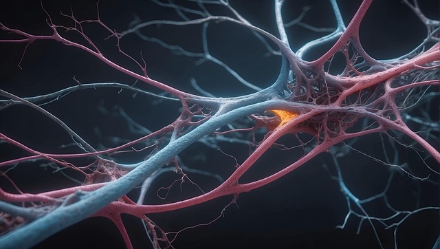
Traumatic Brain and Spinal Injuries (TBI/TSI) require specialized care due to their complex impact on the nervous system, which controls critical bodily functions. Medical imaging techniques like CT scans, MRI, and DTI are vital tools for non-invasive visualization and diagnosis of these injuries. These advanced methods enable healthcare providers to assess damage, identify hidden issues, plan tailored treatments, and monitor recovery, ultimately enhancing patient outcomes through focused interventions on the nervous system using medical imaging for nervous system evaluation.
Traumatic brain injuries (TBI) and spinal cord injuries (SCI) are leading causes of disability globally, emphasizing the need for advanced diagnostic tools. Medical imaging plays a pivotal role in understanding these complex conditions by offering detailed insights into the nervous system. This article explores how cutting-edge imaging techniques, such as CT, MRI, and PET scans, aid in diagnosing TBI and SCI, tracking their progression, and potentially shaping personalized treatment approaches. We also delve into the challenges and future prospects, including ethical considerations, to enhance medical imaging’s impact on patient care.
Understanding Traumatic Brain and Spinal Injuries
Traumatic brain and spinal injuries (TBI and TSI) are severe conditions that demand meticulous care and understanding due to their intricate nature and wide-ranging impacts on the nervous system. These injuries can result from various incidents, such as accidents, falls, or blunt force trauma, leading to life-altering consequences if not promptly addressed. The nervous system, comprising the brain and spinal cord, controls numerous bodily functions, making even minor damage significant.
Medical imaging plays a pivotal role in understanding TBI and TSI by offering non-invasive methods to visualize and diagnose these injuries. Advanced techniques like computed tomography (CT) scans, magnetic resonance imaging (MRI), and diffusion tensor imaging (DTI) enable healthcare professionals to assess the extent of damage, identify hidden lesions or bleeding, and monitor recovery progress. These tools are essential for accurate diagnosis, treatment planning, and long-term management in patients with traumatic brain and spinal injuries, thereby enhancing outcomes through appropriate medical interventions tailored to the specific needs of the nervous system.
– Definition and impact
Traumatic brain and spinal injuries (TBI and TSI) are severe conditions with profound impacts on individuals’ lives and the healthcare system at large. These injuries affect the nervous system, leading to a wide range of physical, cognitive, and emotional impairments. The definition of TBI and TSI encompasses various types of damage, from skull fractures and bleeding to swelling and disruption of neural networks.
Medical imaging plays a pivotal role in understanding and managing these injuries. It provides detailed insights into the structure and function of the nervous system, enabling healthcare professionals to make accurate diagnoses and formulate effective treatment plans. Advanced techniques like computed tomography (CT), magnetic resonance imaging (MRI), and diffusion tensor imaging (DTI) have revolutionized the way we study TBI and TSI, offering a glimpse into the intricate landscape of the brain and spinal cord, and ultimately enhancing patient care in the field of medical imaging for nervous system injuries.
– Common causes and symptoms
Traumatic brain and spinal injuries (TBI and TSI, respectively) are significant global health concerns, often arising from accidents, falls, or violent impacts. The nervous system, comprising the brain and spinal cord, is particularly vulnerable to damage due to its complex structure and sensitivity. Common causes of these injuries include motor vehicle collisions, sports-related traumas, and workplace incidents. Symptoms can vary greatly depending on the severity and location of the injury but may include headaches, dizziness, loss of consciousness, cognitive impairments, sensory disturbances, and motor function deficits.
Medical imaging plays a pivotal role in the diagnosis and understanding of TBI and TSI. Advanced techniques such as computed tomography (CT) scans, magnetic resonance imaging (MRI), and diffusion tensor imaging (DTI) enable healthcare professionals to visualize structural abnormalities, identify damaged areas, and assess the extent of nerve fiber tracts impairment within the nervous system. These imaging tools are crucial for early detection, informed treatment planning, and monitoring patient recovery in the context of traumatic brain and spinal injuries.
The Role of Medical Imaging in Diagnosis
Medical imaging plays a pivotal role in the diagnosis and understanding of traumatic brain and spinal injuries, offering a window into the intricate structures of the nervous system. Techniques such as computed tomography (CT) scans and magnetic resonance imaging (MRI) are indispensable tools for healthcare professionals. CT scans provide rapid, high-resolution cross-sectional images of the brain and spine, enabling doctors to identify acute injuries like haemorrhages, fractures, or bone cracks. MRI, on the other hand, offers a detailed view of soft tissues, helping to detect subtle changes, brain swelling, or nerve damage that may not be apparent on CT scans.
These imaging modalities facilitate accurate assessment, allowing for personalized treatment plans and early intervention. By visually mapping the extent of injuries, medical professionals can better navigate the complex landscape of the nervous system, ultimately improving patient outcomes and recovery prospects for those suffering from traumatic brain or spinal injuries.
Medical imaging plays a pivotal role in understanding and diagnosing traumatic brain and spinal injuries, offering crucial insights into their complex nature. By utilizing advanced techniques, healthcare professionals can accurately assess damage to the nervous system, enabling prompt and effective treatment. This comprehensive approach, centered around medical imaging for nervous system examinations, holds the key to improving outcomes and enhancing recovery for individuals affected by these severe injuries.