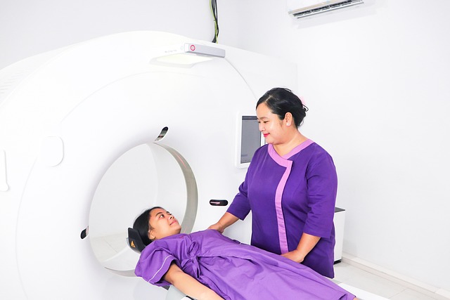
3D nerve mapping leverages advanced neuroimaging scans like MRI and CT to create detailed 3D models of the nervous system. This technique aids surgeons in navigating neural structures, planning precise surgeries, and minimizing damage, thereby improving patient outcomes and recovery times in neurosurgery. Integrating AR/VR and AI algorithms promises further enhancements in surgical accuracy and personalized treatment.
“Revolutionize neurosurgical procedures with 3D nerve mapping, a game-changing technique. This cutting-edge approach transcends traditional 2D methods by providing an intricate, three-dimensional view of complex nerve structures. By ‘unveiling’ these hidden complexities through advanced neuroimaging scans, surgeons gain unparalleled insight during operations.
The benefits are profound: improved accuracy, reduced risks, and enhanced outcomes. This article explores the transformative potential of 3D nerve mapping, delving into its implementation and future prospects in the field of neurosurgery.”
Understanding Nerve Structures: Unveiling Complexity
Understanding nerve structures is a complex task, especially in the context of neurosurgical procedures. The human nervous system is intricate, composed of various types of nerves that form a vast network throughout the body. 3D nerve mapping leverages advanced neuroimaging scans to visualize and analyze these delicate structures in unprecedented detail. By creating three-dimensional models from imaging data, surgeons gain valuable insights into nerve trajectories, branching patterns, and relationships with surrounding tissues. This enhanced understanding allows for more precise surgical planning, minimizing the risk of damage to vital nerves during procedures.
The Role of Neuroimaging Scans in Mapping
Neuroimaging scans play a pivotal role in 3D nerve mapping for neurosurgical procedures. These advanced technologies, such as magnetic resonance imaging (MRI) and computed tomography (CT), provide detailed, high-resolution visualizations of the brain and its intricate neural networks. By combining these scans, surgeons can create comprehensive three-dimensional models that offer unprecedented insight into the complex anatomy of the nervous system.
This precise mapping is invaluable during surgical planning, enabling neurosurgeons to navigate with confidence through critical structures. With accurate 3D representations, they can identify and avoid delicate nerve tracts, blood vessels, and other vital areas, minimizing the risk of damage and enhancing procedural safety. Thus, neuroimaging scans serve as indispensable tools in modern neurosurgery, fostering more precise interventions and ultimately better patient outcomes.
Advantages of 3D Nerve Mapping for Surgeons
3D nerve mapping offers significant advantages for surgeons performing neurosurgical procedures. This advanced technique provides a detailed, three-dimensional view of the nervous system, allowing doctors to accurately identify and navigate delicate neural structures. By integrating data from various neuroimaging scans, such as MRI or CT scans, 3D mapping creates a comprehensive representation of nerves, arteries, and veins in the brain and spinal cord.
This innovative approach enhances surgical precision by enabling surgeons to visualize nerve trajectories, avoid critical structures during dissection, and plan precise incisions. The detailed maps also facilitate better patient outcomes by minimizing damage to surrounding neural tissue, reducing post-operative complications, and potentially improving recovery times.
Implementation and Future Potential in Neurosurgery
The implementation of 3D nerve mapping has revolutionized neurosurgical procedures by providing surgeons with a detailed, precise representation of the intricate neural structures within the brain and spine. This innovative technique integrates advanced neuroimaging scans, such as magnetic resonance imaging (MRI) and computed tomography (CT), to create highly accurate 3D models of nerves, blood vessels, and surrounding tissues. By visualizing these structures in three dimensions, surgeons can better plan complex surgeries, minimize tissue damage, and improve patient outcomes.
Looking ahead, the future potential of 3D nerve mapping in neurosurgery is promising. As technology continues to advance, researchers are exploring ways to enhance the resolution and accuracy of neuroimaging scans, further refining the 3D mapping process. Integration with augmented reality (AR) and virtual reality (VR) platforms could enable surgeons to interact with these detailed models in real-time during procedures, fostering more intuitive and precise surgeries. Additionally, combining 3D nerve mapping with artificial intelligence algorithms holds the key to identifying subtle neural patterns, predicting surgical outcomes, and tailoring treatments to individual patient needs.
3D nerve mapping using advanced neuroimaging scans represents a significant leap forward in neurosurgical procedures. By providing surgeons with detailed, three-dimensional visualizations of complex nerve structures, this technology offers enhanced precision and improved surgical outcomes. As the field continues to evolve, 3D nerve mapping holds immense potential to revolutionize neurosurgery, enabling more effective treatment of conditions that once posed significant challenges. Its implementation promises a future where intricate neural networks are navigated with greater ease, ultimately benefiting patients worldwide.
