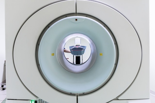
PET scans (Positron Emission Tomography) offer unique insights into brain activity and function not easily accessible via traditional methods like functional nervous system MRI. By tracking metabolic tracers, PET scans highlight areas of high or low brain activity, aiding in the early diagnosis of neurodegenerative diseases like Alzheimer's and Parkinson's. This enables targeted treatment and assessment of treatment effectiveness by monitoring changes in brain metabolism. While structural MRI provides anatomical images, PET scans visualize active brain regions during specific tasks, enhancing the functional perspective in neurological diagnoses. Future advancements in PET technology integrate with functional nervous system MRI, promising revolutionary early disease detection and improved patient outcomes.
“Explore the transformative role of Positron Emission Tomography (PET) scans in unraveling neurological diseases, offering a unique glimpse into brain activity. This article delves into the functional imaging capabilities that set PET apart, allowing for early and accurate detection of disorders affecting the complex neural network.
We compare PET with its prevalent counterpart, MRI, highlighting their distinct strengths. Furthermore, we discuss how PET scans are paving the way for future diagnostic advancements, particularly in understanding the dynamic nature of the nervous system.”
Unveiling Brain Activity: PET Scans' Power
PET scans, or Positron Emission Tomography, are a powerful tool in the field of neurology, offering insights into brain activity and function that other imaging techniques struggle to provide. Unlike traditional MRI (functional nervous system MRI), which primarily visualizes structural changes, PET scans focus on metabolic and functional aspects of the brain. By tracking the flow of tracers, these scans can highlight areas of high or low activity, allowing neurologists to detect abnormalities associated with neurological diseases.
This capability is particularly valuable in diagnosing and understanding conditions like Alzheimer’s disease, Parkinson’s disease, and other neurodegenerative disorders. For instance, PET scans can reveal early signs of brain impairment, identify specific regions affected by the disease, and even measure the effectiveness of treatments by monitoring changes in brain metabolism. By unmasking these hidden dynamics, PET scans play a crucial role in advancing our understanding and treatment of neurological conditions.
Detecting Neurological Disorders: A Functional Approach
Detecting neurological disorders often involves a functional approach, where the focus is on understanding how the brain and nervous system are working rather than just identifying structural abnormalities. This is where PET (Positron Emission Tomography) scans come into play as a powerful tool. Unlike traditional MRI (Magnetic Resonance Imaging), which primarily provides anatomical images, PET scans offer insights into brain function by measuring metabolic activity.
By tracking the flow of tracers, PET scans can highlight areas of the brain that are active during specific tasks or cognitive processes. This functional perspective is particularly valuable in diagnosing neurological diseases like Alzheimer’s or Parkinson’s, where the changes may not be immediately apparent on structural MRI alone. The ability to visualize these subtle shifts in brain function helps neurologists make more accurate diagnoses and plan tailored treatment strategies for each patient.
MRI vs. PET: Comparing Neuroimaging Techniques
In the realm of neuroimaging, Magnetic Resonance Imaging (MRI) and Positron Emission Tomography (PET) scans are two powerful tools used to detect and diagnose neurological diseases. While both offer valuable insights into the brain and nervous system, they do so in distinct ways.
One key difference lies in their approach to visualizing the body’s internal structures. MRI relies on strong magnetic fields and radio waves to create detailed anatomical images of the brain, showcasing its physical characteristics. On the other hand, PET scans utilize radioactive tracers to measure metabolic activity within the brain, providing a window into the functional nervous system. This distinction is crucial when considering specific neurological conditions, as functional MRI (fMRI) can help identify areas of the brain involved in cognitive tasks or disease-related alterations, while traditional MRI captures structural abnormalities.
The Future of Diagnosis: PET's Role in Progress
The future of neurological diagnosis is bright, and Positron Emission Tomography (PET) scans play a pivotal role in this progress. As technology advances, PET becomes increasingly sophisticated, offering more detailed insights into the complex workings of the brain and nervous system. This non-invasive imaging technique has already proven its mettle in detecting various neurological conditions, from Alzheimer’s to Parkinson’s disease, by tracking metabolic activity and identifying abnormal patterns.
With the emergence of advanced instruments and the development of novel radioisotopes, PET scans are evolving beyond their current capabilities. The integration of PET with functional nervous system MRI, for instance, promises even greater accuracy in localizing and characterizing neurological abnormalities. This hybrid approach can potentially revolutionize early disease detection, enabling more effective treatment strategies and improved patient outcomes in the future.
PET (Positron Emission Tomography) scans play a pivotal role in detecting neurological diseases by offering insights into brain activity and function. Their ability to visualize metabolic processes makes them a valuable tool for identifying disorders affecting the functional nervous system. While MRIs provide detailed structural images, PET scans offer a functional approach, highlighting areas of brain activity. As technology advances, PET continues to enhance diagnostic capabilities, especially in combination with functional MRI (fMRI) techniques, paving the way for more accurate and timely interventions in neurology.