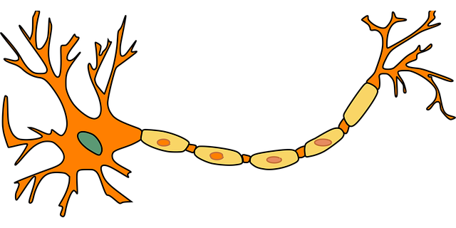
Neurovascular imaging is transforming the assessment and management of traumatic brain and spinal injuries (TBI/TSIs) by providing detailed visualizations of structural changes, inflammation, and vascular alterations. Techniques like Magnetic Resonance Angiography (MRA) and Computed Tomography Angiography (CTA) enable early detection, accurate diagnosis, and personalized treatment plans, ultimately improving patient outcomes and recovery prospects. Ongoing research aims to deepen understanding of these injuries' mechanisms, promising further enhancements in care.
“Unveiling the mysteries of traumatic brain and spinal injuries, medical imaging plays a pivotal role in diagnosis and treatment planning. This comprehensive guide explores cutting-edge techniques, focusing on neurovascular imaging as a game-changer in understanding cerebral trauma. We delve into how advanced technologies offer unprecedented insights into internal injuries, revolutionizing care. From assessing spinal cord damage to visualizing brain abnormalities, these tools are transforming the landscape of medical practice. Discover the future of treatment strategies based on precise, image-guided interventions.”
Neurovascular Imaging: Unlocking Traumatic Brain Insights
Neurovascular imaging has emerged as a powerful tool in unraveling the complexities of traumatic brain and spinal injuries (TBI/TSIs). This advanced technique offers researchers and medical professionals a unique window into the intricate interplay between the nervous system, blood vessels, and their response to trauma. By combining high-resolution imaging with specialized contrast agents, neurovascular imaging can visualize and quantify structural changes, inflammation, and vascular alterations that occur following TBI/TSIs.
This capability is invaluable as it allows for early detection of subtle abnormalities, facilitates accurate diagnosis, and guides personalized treatment strategies. Moreover, ongoing research in this field promises to deepen our understanding of the underlying mechanisms of TBI/TSIs, ultimately leading to improved patient outcomes and enhanced recovery prospects.
Advanced Techniques for Spinal Injury Assessment
Advanced techniques, such as neurovascular imaging, play a pivotal role in assessing and understanding spinal injuries. This innovative approach allows medical professionals to gain detailed insights into the complex interplay between the nervous system and blood vessels within the spine. By employing sophisticated imaging modalities, like magnetic resonance angiography (MRA) and computed tomography angiography (CTA), healthcare providers can visualize spinal cord damage, detect vascular abnormalities, and assess the integrity of neural structures in unprecedented detail.
These advanced imaging methods are particularly valuable for diagnosing and managing traumatic spinal injuries, enabling more accurate treatment planning and improved patient outcomes. With neurovascular imaging, doctors can identify occlusions, aneurysms, or other pathologies that may contribute to neurological deficits, offering a comprehensive understanding of the injury’s extent and potential complications. This, in turn, facilitates targeted interventions and promotes better recovery for individuals affected by these severe injuries.
Enhancing Diagnosis: Visualizing Internal Injuries
Medical imaging plays a pivotal role in enhancing the diagnosis and understanding of traumatic brain and spinal injuries, offering insights into internal damage that might otherwise go undetected. Techniques such as neurovascular imaging have revolutionized the way healthcare professionals assess these complex injuries. By providing detailed visualizations of the brain and spine, these advanced imaging modalities enable doctors to pinpoint exact locations of trauma, identify blood flow disruptions, and detect subtle signs of neurological damage.
Neurovascular imaging techniques, including magnetic resonance angiography (MRA) and computed tomography angiography (CTA), are particularly valuable in revealing injuries to vital structures like blood vessels and neural fibers. This information is crucial for determining the severity of the injury, guiding treatment decisions, and predicting patient outcomes. With these tools, medical teams can offer more precise care, ultimately improving recovery prospects for patients suffering from traumatic brain and spinal injuries.
The Future of Treatment Planning with Medical Imaging
The future of treatment planning for traumatic brain and spinal injuries lies in the enhanced precision offered by medical imaging, particularly neurovascular imaging techniques. These advanced tools provide a detailed glimpse into the intricacies of the brain and spine, allowing healthcare professionals to make more informed decisions. By combining data from various imaging modalities, such as MRI, CT scans, and specialized neurovascular imaging, doctors can accurately identify injuries, assess their severity, and develop personalized treatment strategies.
This precise approach has significant implications for patient outcomes. For instance, neurovascular imaging can help in the early detection of vascular abnormalities or bleeding in the brain, enabling prompt intervention. Moreover, it facilitates a comprehensive understanding of nerve damage, allowing for targeted therapies and potentially reducing long-term complications. With continuous advancements in medical imaging technology, the field holds great promise for improving recovery rates and enhancing the quality of life for individuals affected by traumatic brain and spinal injuries.
Medical imaging plays a pivotal role in understanding and managing traumatic brain and spinal injuries, from providing insights through neurovascular imaging to enhancing diagnosis and treatment planning. Advanced techniques like these are revolutionizing care, ensuring more accurate assessments and ultimately improving patient outcomes. As research progresses, the future of traumatic injury management looks brighter, promising more effective treatments based on detailed visual data.
