MRI and CT scans are powerful tools for nervous system imaging, each with unique strengths. MRI offers superior soft tissue contrast and resolution for detecting delicate nerve structures and subtle damage. CT scans provide rapid, high-resolution cross-sectional images ideal for bone fractures and bleeding assessments. For nerve damage imaging, MRI's detailed visualization makes it the preferred choice due to its ability to capture intricate neural details. The decision between MRI and CT depends on the suspected condition, with each technique contributing uniquely to accurate nervous system evaluation.
When it comes to evaluating the nervous system, choosing the right imaging technique is crucial. This article delves into the comparison between two leading methods: Magnetic Resonance Imaging (MRI) and Computed Tomography (CT) scans. We explore the advantages of MRI for nerve imaging, its superior resolution and contrast sensitivity, making it a game-changer in diagnosing nerve damage. Additionally, we provide insights on when to opt for CT scans, offering a comprehensive guide for healthcare professionals assessing nervous system conditions.
Understanding MRI: Advantages for Nerve Imaging
Magnetic Resonance Imaging (MRI) stands out as a powerful tool for nervous system imaging due to its unique advantages, particularly in detecting delicate structures within the brain and spinal cord. One of its key strengths is its ability to visualize soft tissues with exceptional clarity, which is invaluable when assessing nerve damage or conditions affecting the nervous system.
MRI offers several benefits specifically tailored to nerve imaging. It can provide detailed cross-sectional images, allowing healthcare professionals to identify lesions, inflammations, or anomalies in nerves and surrounding structures. This non-invasive technique also excels in demonstrating minute changes in nerve morphology, making it sensitive enough to detect even subtle forms of nerve damage. Additionally, MRI’s ability to create 3D representations enables comprehensive assessment of the complex architecture of the nervous system.
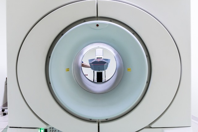
CT Scans: Quick Overview and Their Applications
CT scans, or Computed Tomography, are a rapid and versatile imaging technique that uses X-rays to generate detailed cross-sectional images of the body’s internal structures. This technology has revolutionized medical diagnostics, particularly in emergency settings, due to its speed and accessibility. CT scanners can quickly capture multiple high-resolution images, allowing radiologists to identify and assess various conditions, including nerve damage imaging.
The applications of CT scans are vast, ranging from detecting fractures and tumors to identifying bleeding or infections in the brain and spinal cord. In neurology, CT scans play a crucial role in evaluating structural abnormalities, traumatic injuries, and neurological disorders. They provide valuable information about the brain’s anatomy and can help detect subtle changes related to nerve damage, making them an essential tool for accurate diagnosis and treatment planning.
Comparing Resolution and Contrast Sensitivity
When comparing MRI and CT scans for nervous system imaging, resolution and contrast sensitivity are key factors to consider regarding nerve damage assessment. MRI offers superior soft tissue contrast and spatial resolution, allowing for detailed visualization of intricate neural structures. This makes it particularly advantageous for detecting subtle changes in nerves, such as inflammation or compression, which may indicate nerve damage.
CT scans, on the other hand, provide faster acquisition times and are more cost-effective. While they offer good overall contrast, their lower resolution can limit their ability to distinguish fine details within the nervous system. For comprehensive nerve damage imaging, MRI’s superior resolution and contrast sensitivity make it a preferred choice due to its capability to capture intricate anatomical details that may be missed on CT scans.
Choosing the Best: When to Use Each Method for Nervous System Assessments
When deciding between MRI and CT scans for nervous system assessments, understanding their unique strengths is key. Magnetic Resonance Imaging (MRI) excels in providing detailed, high-resolution images of soft tissues, making it ideal for detecting nerve damage, tumors, or abnormalities within the brain and spinal cord. Its non-ionizing radiation makes MRI a safer choice, especially for repeated scans over time. Conversely, Computed Tomography (CT) scans offer rapid imaging with less patient movement artifacts, allowing for quick assessment of bone fractures or bleeding in the brain. CT is particularly useful when a fast, comprehensive view of the entire nervous system is required, such as in acute traumatic situations.
For nerve damage imaging, MRI often takes the lead due to its superior soft tissue contrast and ability to visualize subtle changes in neural structures. However, if there’s a concern for bone pathology or rapid assessment is critical, CT scans provide valuable insights. The choice ultimately depends on the specific clinical suspicion and patient factors, ensuring the most appropriate method for accurate nervous system evaluation.
When it comes to nervous system imaging, both MRI and CT scans offer valuable insights. MRI excels in detailing soft tissues and providing comprehensive views of neural structures, making it ideal for diagnosing nerve damage and neurological conditions. CT scans, on the other hand, are faster and more accessible, offering high-resolution images of bone structures and blood vessels, which can aid in identifying associated nervous system pathologies. Choosing between them depends on specific clinical needs, patient factors, and the type of nerve damage imaging required. In many cases, a combined approach using both modalities can provide the most comprehensive assessment.